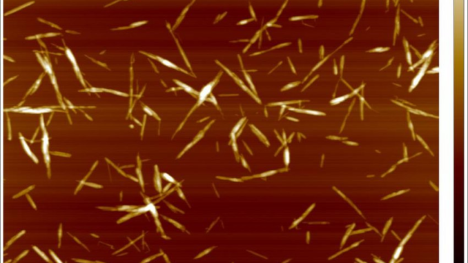
Image processing has widened the scientific scope dramatically since its creation, and can now be used to assess live tissue, single cells and molecules and for bio-chemical analysis. This is understandably having a huge impact on both modern medicine and the pharmaceutical industry, as well as broadening our understanding of live cells.
But as you will know, in order to keep abreast of the latest research, you’ll also need to invest in the latest technology. When it comes to assessing samples, the microscope you use is crucial to the accuracy of your research, so you need to make sure you’re choosing the right AFM for your laboratory or research facility and getting the best equipment for your money.
To start with, you’ll need to assess your options online by researching some AFM providers. It’s a good idea to consult other scientists on trusted sites or forums to get an idea about the best company from which to purchase your technology. You can usually find customer testimonials or complaints on review sites, too.
Be sure to check out the credentials of your chosen supplier. Although some companies are more affordable than others, this kind of technology doesn’t come cheap – so make sure you’re getting the best possible model for your money, and find out what your options are if the machine gets damaged or stolen.
It’s also important to choose a company who can offer you training alongside the setup of your new model. This training shouldn’t last longer than a couple of hours, but will give you and your team a comprehensive overview so you can start conducting more advanced research straight away. The company should also be available for after-sales support as well.
In order to keep set-up time to a minimum and ensure productivity across the board, you’ll need to find an AFM that is easy to use, even for entry level researchers. Look out for providers advertising user-friendly models and automated imaging software, so you won’t have to waste time figuring out how the technology works.
In terms of technical features, the latest imaging technology should be set up for SICM – Scanning Ion Conductance Microscopy. This will allow you to analyse samples that are completely immersed in liquid, and will in turn provide the most accurate results possible. SICM models will typically use glass nanopipettes filled with an electrolyte that acts as an ion sensor.
SICM models can image all cells, even soft cells such as neuron cells and live tissue – something that hasn’t been possible until very recently, and is simply not an option when using other microscopy techniques. SICM can even image suspended networks of neurons, unlike other microscopes.
The most recent models will also offer no force, non-contact image processing, so it’s important to look out for these features when you’re researching AFMs. Instead of making contact with the sample (and subsequently interfering with the results), the pipette of a recent SICM model will maintain considerable distance from the sample itself.
In SICM, a sensor measures the current flow from the ionic current that flows from the pipette through the solution surrounding the sample, which then decreases the distance between the pipette and the sample. This distance is then monitored in order to obtain the topology of the artefact being analysed.
Be sure to get your new model insured. Whether you are able to do this through the company you purchase it from, or source it externally, it’s vital that you protect your company from any unexpected costs and interrupted research if the model gets damaged or stops working.
Proudly WWW.PONIREVO.COM
by Stevens P


